Continued Success for EVF HOW: the 5th Workshop is Organized in Cyprus 2014
2 Helsingborg, Sweden
3 Nicosia, Cyprus
The concept of a hands-on workshop on venous disease was introduced during the “The Arctic Fjords Conference and Workshops on Chronic Venous Disease” in 2007. The workshop was well received. One of the major objectives of the European Venous Forum (EVF) is to develop and provide education within the venous field. Since there was a lack of practical courses on the clinical management of chronic and acute venous disease, the decision was to further develop the workshop concept under the auspices of EVF. In October 2010, the 1st EVF HOW (European Venous Forum Hands-on Workshop on Venous Disease) was organized and it proved to be successful. The goal is not only to provide understanding of modern practical management, but also for the delegates to learn hands-on individual procedures to treat venous disease. An integral partner in this effort is the providers of different devices, sclerosing agents, stockings, bandages, ulcer care material, ultrasound scanning machines, etc. The objectives of the EVF HOW are impossible to fulfill without this partnership. The workshop targets those who want an introduction to or need an update on the management of venous disease. Its intention is to provide a broad teaching on all aspects of acute and chronic venous diseases. EVF HOW is open to all specialty physicians, including physicians in training. The 5th EVF HOW will take place at the Grand Resort, Limassol, Cyprus, between October 30th and November 1st, 2014. As only 100 participants are accepted on a “first-come, first-served” basis, it is recommended to register early to ensure a place. Please contact Anne Taft, Administrative Director, European Venous Forum; tel/fax +44 (0)20 8575 7044; email admin@europeanvenousforum.org. More information is available at www.europeanvenousforum.org.
The Objectives
To educate, train, and update learners (the delegates) on the current clinical management of patients with venous disease by close informal interaction with venous experts during lectures, case discussions, and hands-on activities in small groups.
At the end of this course, the learner (delegate) should be able to:
1. I dentify venous disease in patients.
2. A pply appropriate venous investigations.
3. Construct a plan for management.
4. U nderstand different interventional procedures.
5. Successfully incorporate treatment of venous patients in his/her practice.
6. R ealize when to refer a patient for expert care.
The Format
The EVF HOW mission statement is to provide “education and hands-on practice for the benefit of patients with venous disease.” The instruction of learning has been structured from the start on a few principles, which are important for the success of the workshop. The number of learners are limited (max 100 delegates) to facilitate interaction between instructors and delegates and thus the faculty/learner ratio is high (1/3). The hands-on sessions are truly hands-on for the delegates, and are not small lectures or only a demonstration of procedures. All learning sessions are informal in a relaxed setting to allow uninhibited communication between delegates, faculty members, and industry representatives. Plenty of time is set aside for discussion with the greatest interaction occurring at the workshop stations. The learners are encouraged to bring their own cases for presentation and discussion. There is no exhibition or parallel activity.
The format of the EVF HOW in 2014 will be similar to previous years including formal lectures, case discussions, and live demonstrations on duplex scanning covering acute and chronic venous diseases. The focus will be on handson training on procedures and devices. Faculty members in collaboration with the industry experts will instruct at 20 to 24 workshop stations. The delegates will attend each workshop station for 30 min in small groups (4 to 5 delegates), which will give each participant time to try out devices, practice with the bandages, etc.
Learning Enhanced by New Website
Last year the EVF VIP (European Venous Forum Venous Interactive Portfolio) was introduced. This is a webbased portfolio where each delegate has access to the presentations, important references and guidelines, the case reports, videos of procedures, supplementary information about the workshop stations, and other study material. Participation in the EVF HOW will provide access to this longterm portfolio. The long-term availability to the website gives the learners a possibility to go back, reinforce, and enhance their learning experience. The response after the EVF HOW in Stockholm 2013 was very positive. A majority of learners used the website before the start of the workshop (86%) and as many as 48% accessed it during the workshop. All participants fully or partially agreed that the EVF VIP was a valuable supporting tool especially having access to the presentations, availability of references and guidelines in pdf format, and a user-friendly overview of the program were most appreciated.
4th EVF HOW Stockholm, 2013
The 4th EVF HOW was organized at the Marina Tower, Elite hotel in Stockholm, Sweden for three days from October 31st to November 2nd last year. The primary reason given for attendance by the delegates was to update overall knowledge about venous disease and its treatment (78%), to be introduced to venous disease (13%), and to learn particular techniques (9%). In an assessment after the course, the overwhelming majority of the delegates indicated that these goals were achieved (97%). The learners felt that the workshop stations achieved all or most stated goals between 95% and 100% at each station. Overall, the learners felt that their expectations were met (98%) and that the workshop would impact and change their practice in the future (92%). More than half of the delegates were vascular surgeons (67%) followed by other specialties such as general surgeons (11%), phlebologists (8%), interventional radiologists (6%), angiologists, cardiologists, and dermatologists. The EVF HOW was created mainly for Europe; 55% and 22% are from Western and Eastern Europe, respectively. The workshop has attracted international attention with representation from all over the world including 11% from the Middle East, 4% from Asia, and 8% from Africa, South America, USA, and Australia. Thus, the concept of the EVF HOW (Hands-on Workshop on Venous Disease) has been internationally well received.
The Program of EVF HOW 2013
The instruction at the 4th Hands-on Workshop on Venous Disease in 2013, was provided by an international faculty with 30 experts from Europe and the USA. They not only give presentations, but also actively discussed case presentations and were an integral part of the workshop giving practical tips and tricks from their own experience. The clinical input by the faculty members balanced the specific device information presented by the industry representatives.
Presentations
The presentations spanned the following subjects:
• Basic principles of venous pathophysiology, accuracy of tests, and classification and assessment of treatment outcomes.
• Treatment of varicose veins conservatively with drugs and compression, with invasive procedures such as open surgery or saphenous ablation with laser, radiofrequency, foam sclerotherapy, steam, and pharmacomechanical means, and with techniques preserving the saphenous vein. After intense discussion, Professor Andrew Bradbury tried to make sense of it all. The controversies of the perforators were elucidated and interventions for recurrent or residual varicose veins (PREVAIT , Presence of varices after intervention) were outlined.
• Guidelines for prevention and treatment of venous thromboembolism (VT E) and superficial thrombophlebitis (SVT ).
• Treatment of acute VTE with traditional conservative measures, new oral anticoagulants, catheter-directed thrombolysis, and pharmacomechanical thrombectomy was described and the outcomes presented, and the role of inferior vena cava (IVC) filters was presented.
• Pelvic congestive syndrome.
• Diagnosis and treatment of chronic venous insufficiency using a sequential treatment plan was presented including compression treatment, the role of fasciotomy in legs with increased compartment pressure, treatment of deep venous obstruction, and the role of valve reconstruction in limbs with primary deep venous reflux or postthrombotic disease including the use of the Vedensky spiral.
Case reports
There were 18 interesting cases presented. This year the case reports were imbedded among the formal presentations, which appeared to significantly increase the participation. Only five cases were brought by the delegates for discussion and the remaining were provided by the faculty. In the future, we hope that more learners will bring their own cases. Each case was presented in stages and the moderator encouraged the delegates to join in at all stages, which lead to lively discussions. There was a wide range of cases illustrating the previously given lectures: From varicose veins to acute iliofemoral deep venous thrombosis (DVT ); from chronic outflow obstruction to ovarian venous reflux. Hands-on workshops As previously emphasized, this component of the EVF HOW is the most important. The function of the device or the method presented at each workshop station was explained in detail by the industry expert. Its role in the treatment of venous disease and personal clinical tips and tricks were highlighted by the faculty member. Each learner trained hands-on under expert supervision after a short demonstration.
Workshop 1
The learners performed live imaging in patients with different types of vein pathologies. Drs Anders Holmberg and Lena Blomgren had collected numerous patients from their practice, representing a variety of disease. The aim was two-fold. First, the learner should be able to position the patient properly, use appropriate transducers, know imaging principles, and optimize the image. Second, the learners should be able to identify acute and chronic disease, reflux, obstruction, and pathology surrounding the vessels. Station 1: Lower limb with normal findings (Siemens; faculty Ragnhild Östmyren/Andrew Nicolaides) (Figure 1).
Station 2: A bdominal and pelvic vein investigation (GE Healthcare; faculty: Nicos Labropoulos/Stefan Rosfors).
Station 3: Lower limb with superficial reflux (Philips; faculty: Lena Blomgren/Lena Persson).
Station 4: Lower limb with deep incompetence (Zonare; faculty: Niki Georgiou/Kent Lund).
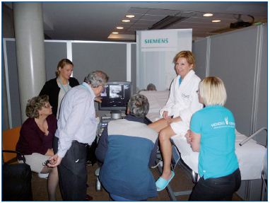
Figure 1. Learner practicing ultrasound scanning of the lower
limb.
Workshop 2
Station 1: Venous stenting. Placement of the Veniti Vici venous stent was practiced by each learner in a specially designed venous tubular model replicating the ilio-caval vein segment (Veniti; faculty: Marzia Lugli/Oscar Maleti) (Figure 2).
Station 2: IVC filter placement and retrieval was practiced in a tube model (Cook Medical; faculty: Anthony Gasparis/Evgeny Shaydakov). Station 3: Compression therapy/ulcer care. The delegate learned how to choose and apply the appropriate wound dressing for a venous ulcer and how to measure a leg and apply the appropriate stocking (BSN/Jobst; faculty: Sylvain Chastenet) (Figure 3).
Station 4: Early clot removal with the Trellis™ – Peripheral Infusion system. After a brief demonstration of the technique, the Trellis device was prepared, inserted, and used in a model by the learners (Covidien; faculty: Gerard O’Sullivan). measuring the working pressure with these stockings by using a Picopress device. (Bauerfeind; faculty: Michel Perrin).
Workshop 3
Station 1: Laser saphenous ablation. After longitudinal and transverse access to the vein under ultrasound guidance, saphenous laser ablation was practiced on a phantom leg using a radial fiber with a 1470 nm laser generator. Tips and tricks were given and how to decide the dosage of energy was practiced. (Biolitec; faculty: Anders Holmberg).
Station 2: Stocking. The learners practiced choosing a correct medical compression stocking (MCS) by measurement and applying long- and shortstretched MCS with and without fitting aid and measuring the working pressure with these stockings by using a Picopress device. (Bauerfeind; faculty: Michel Perrin).
Station 3: Steam saphenous ablation. The Veni RF Plus steam device was used. The physical effect of the technique was explained followed by demonstration of the setup of the generator and catheter. Steam ablation following catheterization was practiced individually by the learners using a vein model (Veniti; faculty: Marianne De Maeseneer).
Station 4: Foam sclerotherapy. The learners made foam using sodium tetradecyl sulphate (STD) and discussed different treatment plans according to ultrasound scanning results. This was followed by ultrasoundguided cannulation and injection of foam in a phantom leg. The appropriate compression bandage following foam sclerotherapy was placed on each other (STD Pharma; faculty: Andrew Bradbury and Gareth Bate).
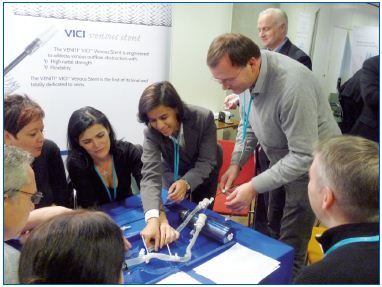
Figure 2. Learner practicing stent placement in a tubular
model.
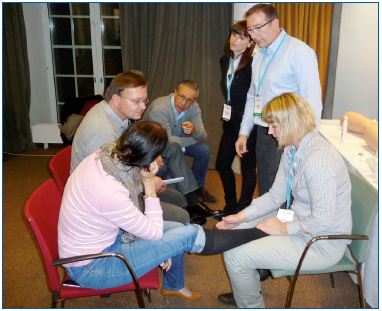
Figure 3. Learners practicing application of stocking.
Workshop 4
Station 1: Ovarian vein embolization was practiced by the learners in a specially designed tubular venous model (Cook Medical; faculty: Jan Engström).
Station 2: R F saphenous ablation. Saphenous radiofrequency ablation using the ClosureFAST catheter was practiced by the learner including how to accurately place the catheter tip at the saphenofemoral confluence and to sequentially position the catheter (Covidien; faculty: Lars Rasmussen/ Athanasios Giannoukas).
Station 3: Bandage. Strong short-stretch compression bandage was applied by each learner, subbandage pressure measurements were monitored, and the learners were made aware of what a correctly applied bandage on their own leg feels like (Lohmann & Rauscher; faculty: Hugo Partsch/Giovanni Mosti) (Figure 4).
Station 4: T he concept of medi-Circaid inelastic compression device was explained and the device applied by each delegate (medi; faculty: Sandra Shaw).
Workshop 5
Station 1: Early clot removal with continuous catheter directed thrombolysis (CDT ) using the Unifuse CDT catheter. The learners individually practiced insertion of the catheter and the occluding ball wire and were instructed on how to set up a protocol for CDT treatment (Angiodynamics; faculty: Niels Baekgaard)
Station 2: V enous stenting. The learners practiced deployment of a Wallstent in a tubular model at the IVC confluence and distally in the iliofemoral vein. The specific properties of a braided stent were demonstrated (Boston Scientific; faculty: Antonio Rosales).
Station 3: Laser saphenous ablation. The learner practice saphenous ablation using the KLS Martin endovenous laser including planning of adequate dosage, selection of the correct treatment set, access to the vein, precise placement of the laser fiber intraluminally and correct application of the laser energy on a phantom limb (KLS Martin Group; faculty: Zbigniew Rybak) (Figure 5).
Station 4: Foam sclerotherapy. The learner practiced how to produce foam with Aethoxysklerol® and the EasyFoam® Kit. Cannulation of larger veins and the injection of tiny spider veins were practiced using the phantoms and ultrasound or the portable vein finder Veinlite LED®. Tips and tricks for optimal results were pointed out (Kreussler; faculty: Eberhard Rabe).
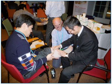
Figure 4. Learners placing bandages on each other.
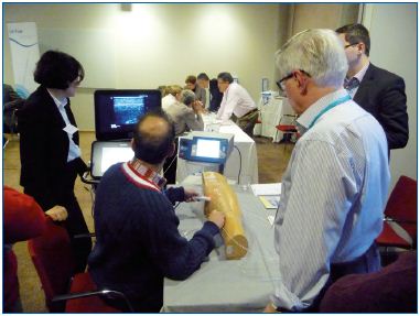
Figure 5. Learner practicing saphenous ablation on a phantom
leg.
Workshop 6
Station 1: Insertion of the Crux® Vena Cava Filter was performed by each delegate using a computerized Mentice simulator mimicking an angioroom set up (Volcano/Mentice; faculty: Lars Lönn) (Figure 6).
Station 2: N onthermal saphenous ablation. After being informed about the technical aspects of the nonthermal Clarivein ablation technique, the learners practiced placement of the device by ultrasound guidance on a blue phantom limb and performed a mock treatment (Vascular Insights; faculty: Steve Elias).
Station 3: Stockings. The learners practiced measurement of the leg dimensions and application of a stocking on each other, learned how to prescribe an appropriate stocking, and experienced the difference in material use in manufacturing of stockings (Sigvaris; faculty: Olle Nelzén).
Station 4: Intravascular Ultrasound (IVUS). The learners familiarized themselves with the IVUS tower and the corresponding catheters. Case studies of procedures using IVUS video-loop recordings were reviewed to demonstrate differences in venogram and IVUS images during treatment of patients with obstruction (Volcano; faculty: Jan Christenson).
Figure 6. Learners using a computerized simulator for IVC
placement.
Evaluation and future meeting
The 4th EVF HOW in 2013 was appreciated by the delegates, faculty members, and industry representatives. They all enjoyed the learning sessions because of the informal and close interaction. A pre- and post-MCQ test was performed and showed an average improvement of 40% after theworkshop, which clearly indicated that the workshop had the intended impact on learning.
The 5th EVF HOW will have similar structure as outlined above. It will be limited to 3 days and will require full attendance. If a participant wants to include some sightseeing, we suggest that this is arranged by prolonging the stay in Cyprus. There will be no sightseeing arranged and no program for accompanying persons. The organizers hope to further improve the quality of the content by taking into account constructive criticism by previous learners. More case reports brought by the learners are necessary and encouraged. Perhaps it will be possible to further develop the EVF VIP website and create personal portfolios with additional study material.
Corresponding author